How Osteoporosis Looks Like in CT Scan Images: An Alternative Osteoporosis Assessment
Keywords:
CT-scan, Osteoporosis, Osteopenia, Bone Quality, Attribute, ImagingAbstract
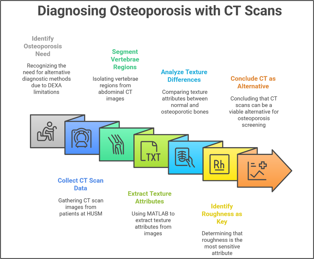
Osteoporosis is a silent degenerative disease that commonly affects the elderly, leading to decreased bone density and increased risk of fractures. Dual-Energy X-ray Absorptiometry (DEXA) is the standard tool for assessing Bone Mineral Density (BMD), but its limited availability and high cost in many healthcare facilities, especially in developing countries like Indonesia, necessitate alternative diagnostic methods. This study aims to assess the potential use of Computed Tomography (CT) scan images as a substitute for DEXA in detecting osteoporosis. The objective is to analyze bone texture attributes from CT images to differentiate between normal and osteoporotic bone structures. The method involved collecting CT scan data from patients at Hospital Universiti Sains Malaysia (HUSM). The vertebrae regions were segmented from the abdominal CT images using image processing techniques to isolate bone tissue. Four image-derived texture attributes—Roughness, Contrast, Greyscale, and Phase—were then extracted using MATLAB-based analysis. The results showed that osteoporotic bones had higher values in Roughness and Contrast, and lower values in Greyscale and Phase compared to normal bones. Among these, Roughness was identified as the most sensitive attribute in detecting changes associated with osteoporosis. These findings indicate that CT scan images, when analyzed through proper segmentation and texture evaluation, have the potential to serve as a viable alternative for osteoporosis screening, particularly in settings where DEXA is unavailable.
Downloads
References
BPS. Publikasi [Internet]. 2024. Available from: https://www.bps.go.id/id/publication
Sözen T, Özışık L, Başaran NÇ. An overview and management of osteoporosis. Eur J Rheumatol. 2016;4(1):46.
Choi YJ. Dual-energy X-ray absorptiometry: beyond bone mineral density determination. Endocrinol Metab. 2016;31(1):25.
Sardjito Hospital. Osteoporosis [Internet]. 2019. Available from: https://sardjito.co.id/2019/09/30/osteoprosis/
Brown JP, Engelke K, Keaveny TM, Chines A, Chapurlat R, Foldes AJ, et al. Romosozumab improves lumbar spine bone mass and bone strength parameters relative to alendronate in postmenopausal women: results from the Active‐Controlled Fracture Study in Postmenopausal Women With Osteoporosis at High Risk (ARCH) trial. J Bone Miner Res. 2021;36(11):2139–52.
Akkawi I, Zmerly H. Osteoporosis: current concepts. Joints. 2018;6(02):122–7.
Haseltine KN, Chukir T, Smith PJ, Jacob JT, Bilezikian JP, Farooki A. Bone mineral density: clinical relevance and quantitative assessment. J Nucl Med. 2021;62(4):446–54.
Engelke K, Chaudry O, Bartenschlager S. Opportunistic screening techniques for analysis of CT scans. Curr Osteoporos Rep. 2023;21(1):65–76.
Pickhardt PJ, Pooler BD, Lauder T, del Rio AM, Bruce RJ, Binkley N. Opportunistic screening for osteoporosis using abdominal computed tomography scans obtained for other indications. Ann Intern Med. 2013;158(8):588–95.
Genisa M, Abdullah JY, Yusoff BMD, Arief EM, Hermana M, Utomo CP. Adopting signal processing technique for osteoporosis detection based on CT scan image. Appl Sci. 2023;13(8):5094.
Soles GLS, Ferguson TA. Fragility fractures of the pelvis. Curr Rev Musculoskelet Med. 2012;5:222–8.
Oberkircher L, Ruchholtz S, Rommens PM, Hofmann A, Bücking B, Krüger A. Osteoporotic pelvic fractures. Dtsch Arztebl Int. 2018;115(5):70.
Hendrickson NR, Pickhardt PJ, Del Rio AM, Rosas HG, Anderson PA. Bone mineral density T-scores derived from CT attenuation numbers (Hounsfield units): clinical utility and correlation with dual-energy X-ray absorptiometry. Iowa Orthop J. 2018;38:25.
Genisa M, Rajion ZA, Mohamad D, Pohchi A, Kadir MRA, Shuib S. Effect of Different Angle Scanning on Density Estimation Based on Hounsfield Unit on CT and CBCT. Sains Malaysiana. 2015;44(9):1331–7.
Van Oostwaard M. Osteoporosis and the nature of fragility fracture: an overview. Fragility Fract Nurs Holist care Manag orthogeriatric patient. 2018;1–13.
Kim MW, Noh YM, Huh JW, Seo HE, Lee DH. Comparative Analysis of CT Texture in Lumbar and Femur and Its Correlation with Bone Mineral Density and Content over Time: An Exploratory Study. Diagnostics. 2023;13(23):3588.
Zou D, Li W, Deng C, Du G, Xu N. The use of CT Hounsfield unit values to identify the undiagnosed spinal osteoporosis in patients with lumbar degenerative diseases. Eur Spine J. 2019;28:1758–66.
Kawashima Y, Fujita A, Buch K, Li B, Qureshi MM, Chapman MN, et al. Using texture analysis of head CT images to differentiate osteoporosis from normal bone density. Eur J Radiol. 2019;116:212–8.
Published
How to Cite
Issue
Section
Copyright (c) 2025 Maya Genisa, Johari Yap Abdullah, Bazli Bin MD Yusoff, Aryenti Aryenti, Juniarti Juniarti

This work is licensed under a Creative Commons Attribution-NonCommercial-ShareAlike 4.0 International License.
Similar Articles
- Nur Azmi, Ayu Prawesti Priambodo, Aan Nuraeni, Analysis of Handoff Communication Using SBAR (Situation, Background, Assessment, Recommendation) in Emergency Department and Intensive Care Unit: A Scoping Review , Journal of Health and Nutrition Research: Vol. 4 No. 2 (2025)
You may also start an advanced similarity search for this article.